Best Centre For Prenatal Imaging And Diagnostic Scans In Mumbai
Let’s begin the journey to motherhood with clarity and care
Best Centre For Prenatal Imaging And Diagnostic Scans In Mumbai
Prenatal Imaging And Diagnostic Scans
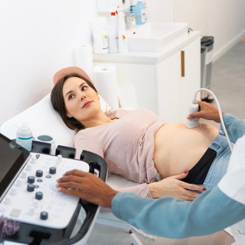
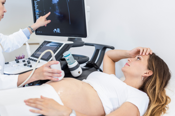
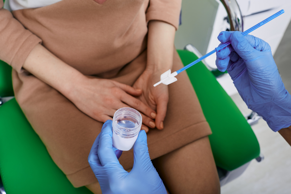
Prenatal Imaging and Diagnostic Scans refer to medical tests and imaging techniques used during pregnancy to monitor the health and development of the fetus, as well as to assess the well-being of the mother. In this regard, we are preferred by those who need the best centre for prenatal imaging and diagnostic scans in Mumbai due to our professional staff, advanced machines, expertise and accuracy.
50k+
Ultrasound Scans
15k+
Detailed Anomaly Scans
1k+
3D Scans

1. Early Pregnancy Viability Scan
An early pregnancy scan is the first scan to have a look at your baby, mostly chosen by couples for reassurance. It helps ensure that it is not an ectopic pregnancy, and there can be many other reasons to get it done.
When Is It Performed?
- It is performed between 6 and 10 weeks of pregnancy in the first trimester.
- It helps find whether you are expecting twins or a singleton and predict the due date.
- It is also performed when assurance of the regular beating of the baby’s heartbeat is needed.
2. First Trimester (NT-NB) Scan
A Nuchal Translucency/Nasal Bone (NT-TB) Scan helps assess the risk of the baby having specific chromosomal abnormalities. The NT scan measures the space filled with fluid behind the neck of your unborn baby, while the NB scan helps determine the absence or presence of the nasal bone, which can indicate potential chromosomal issues.
When Is It Performed?
- It is performed between 11 and 14 weeks of pregnancy in the first trimester.
- It is recommended when a woman’s age, family history, lifestyle, etc, can increase risks.
- It can also be recommended if the previous tests show potential risks.

3. Early Anomaly Scan
The Early Anomaly Scan is important and helps determine if the developing baby has significant physical anomalies. These anomalies can be detected in the limbs, spine and other vital organs.
When is it done?
- It is the second-trimester scan performed between 16 and 18 weeks of pregnancy.
- It helps determine the correct development of the facial characteristics and organs.
- It can be performed to identify chromosomal abnormalities.
4. Detailed Anomaly Scan
The Detailed Anomaly Scan is similar to a standard ultrasound scan but provides detailed information. Here, fetal anatomy is assessed to ensure that the baby develops normally, especially by measuring the parts of the body to see how well it is growing.
When Is It Performed?
- It is a second-trimester scan performed between 18 and 23 weeks.
- It helps determine the presence of Spina Bifida (Open Spinal Cord).
- It is also done when special attention is needed for the baby’s brain, face and other vital organs.
5. Fetal Echocardiography
This painless ultrasound test helps see the baby’s heart structure and its functioning. If an abnormality is detected during the routine scan or if the routine obstetrical ultrasound did not clearly show the baby’s heart, Fetal Echocardiography can help.
When Is It Performed?
- It is performed between 18 to 24 weeks in the second trimester.
- It can be performed if there is a related family history or the baby has a genetic disorder.
- It is also needed if the mother has a condition that increases the risk to the baby’s heart.
6. Fetal Neurosonography
Fetal Neurosonography is a specialised ultrasound technique focused on assessing the fetal brain and spinal cord to detect potential neurological abnormalities.
When Is It Performed?
- It is performed during the second trimester, between 18 and 24 weeks of gestation.
- It can be recommended if an assessment of structural abnormalities is needed.
- It can be needed to detect various conditions affecting the brain and the spine.


7. Fetal Well-Being Scan
During this scan, the baby’s measurement is taken to estimate fetal weight, assess amniotic fluid levels and fetal movement. Baby’s size may vary due to various factors. Thus, the scan is recommended to ensure that the baby grows well without complications.
When Is It Performed?
- It is performed between 24 and 40 weeks of pregnancy in the second and third trimesters.
- It is recommended when fetal movements such as breathing and stretching are to be observed.
- It can also be recommended to determine the position of the baby.
8. Fetal Colour Doppler + Biophysical Profile
Unlike standard Doppler scans, Fetal Colour Doppler converts sound waves into colour images to provide a clearer view of blood flow in the placenta and uterus. On the other hand, the Biophysical Profile (BPP) helps assess the health of the fetus via fetal heart rate monitoring and ultrasound.
When Is It Performed?
- It is performed between 28 and 32 weeks in the third trimester.
- It is needed to assess the blood flow, heartbeat and overall well-being of the baby.
- It is also needed to monitor uneven growth patterns in twins or multiple fetuses.


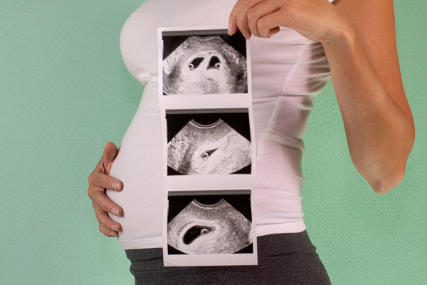
9. Fetal 3D/4D Scans
The 3D scans are created by merging the 2D pictures, and the 4D scans are more like real-time photographs of your unborn baby. The 3D scans help diagnose the neural defects, fetal face defects and view the fetal heart structures, while the 4D scans help examine the fetal heartbeat and overall well-being and growth of the baby.
When Are They Performed?
- These scans are performed between 23 and 36 weeks of pregnancy in the second and early third trimester.
- They can be recommended when advanced ultrasound technology for detailed imaging is needed.
- They also help in capturing a baby’s movements, like yawning or stretching.
10. Fetal 3D/4D Mould Prints
These prints capture detailed facial and body details from inside the womb and show the baby’s facial expressions, body movements and overall structure. They can complement the 2D scans but not replace them for routine assessments.
How Do They Help?
- They help in counselling and planning for post-medical treatments.
- They provide better visualisation of structural abnormalities.
- Overall, they help determine the well-being of the baby.

11. Cervical Screening
Cervical Screening is performed to check the health of the cervix. It is not performed to test for cancer, but rather helps prevent cancer by looking for abnormal changes. If abnormal cells are found, they can be treated on time, thus, it is an important test, yet optional.
How Does It Help?
- Women can opt for this test from the age of 21.
- It can help check for high-risk types of HPV.
- It can be recommended if the person has a family history of related concerns.
12. Fetal MCA-PSV Monitoring
MCA-PSV Test (Middle Cerebral Artery Peak Systolic Velocity Test) is a non-invasive Doppler ultrasound technique used to measure the peak blood flow velocity in the fetal middle cerebral artery (MCA).
When Is It Performed?
- It is ideal to be performed between 25 and 35 weeks of gestation in the second and third trimesters.
- This test is primarily used to detect fetal anaemia, especially in pregnancies at risk.
- It helps decide if early treatment or delivery is necessary.


13. Placenta Accreta Spectrum
Placenta accreta is a serious pregnancy condition where the placenta grows too deeply into the uterus’ wall. In a normal pregnancy, the placenta detaches from the uterus after the baby is born. However, in placenta accreta, it remains firmly attached, which can lead to severe bleeding during or after delivery.
When Does It Get Diagnosed
- It is usually diagnosed during pregnancy via ultrasound or MRI.
- Mostly, it gets diagnosed in the second and third trimesters.
- In some cases, it is only discovered after delivery when the placenta fails to detach normally.
Let’s Track How Your Little One Is Growing
FAQs
How to prevent placenta accreta?
Placenta accreta cannot be prevented by the mother. The risk of having placenta accreta increases if the mother has had a placental disorder or multiple C-sections. However, it can be diagnosed early via prenatal ultrasound to be prepared for the required procedures. In this regard, we provide guidance and care at various stages in your pregnancy. Thus, our Bliss Fetal Medicine Centre can turn out to be ideal for those who need a trustworthy centre for sonography in Vasai Virar or an early pregnancy scan in Vasai Virar, Mumbai.
Can ultrasound detect due date?
Yes, ultrasound helps detect due date, and an Early Pregnancy Viability Scan is performed for that in the 1st trimester between the 7th and 12th week. So, if you are looking for ultrasound / sonography centres in Vasai Virar, Mumbai, Bliss Fetal Medicine Centre can be ideal because we are known for performing precise early pregnancy scans and providing related counselling for your healthy pregnancy journey.
Should my bladder be full of water before appearing for every ultrasound scan?
Having a full or empty bladder depends on the type of ultrasound scan. For an Early Pregnancy Scan, Third Trimester Growth and Doppler Scan and Fetal Neurosonography, the bladder should be empty. For an NT Scan, Anomaly Scan and Fetal Echocardiography, the bladder should be full. Meanwhile, for a 3D/4D Scan, it depends on the month of pregnancy when it’s done. So, it is always best to follow an expert’s advice. If you are looking for prenatal imaging and diagnostic scans or a sonography clinic in Mumbai, our Fetal Medicine Centre can stand as a prominent choice where you can discuss your queries with our specialist and be prepared for a scan accordingly.
What if I miss an early pregnancy scan?
If you miss an early pregnancy scan, contact your obstetrician and schedule one as soon as possible. It is because this type of scan helps confirm pregnancy and reveals if there are multiple embryos. Similarly, it helps detect some anomalies so that they can be worked upon from an early stage. In this regard, our centre provides early pregnancy scan in Vasai Virar, Mumbai, with required guidance and counselling so that you can ensure complete care for your unborn little one.
Should I bring any documents for the scans?
Yes, a photo ID proof (Aadhar Card) is necessary for a pregnancy ultrasound at our centre. Similarly, a doctor’s paper with a sign and a stamp is compulsory. It is how we ensure that we follow proper ethical guidelines, and that’s why we are preferred by those looking for the best sonography centre in Vasai, Mumbai.
Can Fetal 3D/4D Scans analyse birth defects?
Yes, Fetal 3D/4D scans can help analyse certain birth defects. A 4D and 3D sonography in pregnancy is useful in detecting limb abnormalities, neural tube defects and some congenital heart issues. Also, it analyses any facial clefts (Cleft Lip). If you are looking for a sonography clinic in Mumbai or a centre for sonography in Vasai Virar, our Fetal Medicine Centre stands apart because we use these advanced imaging techniques for a more accurate assessment of fetal health.
prenatal imaging and diagnostic scans
prenatal imaging and diagnostic scans
prenatal imaging and diagnostic scans
prenatal imaging and diagnostic scans
prenatal imaging and diagnostic scans
prenatal imaging and diagnostic scans
prenatal imaging and diagnostic scans
prenatal imaging and diagnostic scans
prenatal imaging and diagnostic scans
prenatal imaging and diagnostic scans
prenatal imaging and diagnostic scans
prenatal imaging and diagnostic scans
prenatal imaging and diagnostic scans
prenatal imaging and diagnostic scans
prenatal imaging and diagnostic scans
prenatal imaging and diagnostic scans
prenatal imaging and diagnostic scans
prenatal imaging and diagnostic scans